Evaluation of morphological characteristics of retinal vessels

The accurate and timely monitoring of hypertension-related diseases is important for a population screening and follow up, to prevent the onset and to assure proper treatment. The evaluation of morphological characteristics of small resistance arteries in human beings in not easy. The gold standard is generally considered the evaluation of the media to lumen ratio of subcutaneous small vessels obtained by local biopsies and measured by wire or pressure micromyography. 

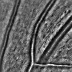
However, non-invasive techniques for evaluation of retinal arterioles were proposed, in particular two approaches seem to provide interesting information: scanning laser Doppler flowmetry and adaptive optics; both of them provide an estimation of the wall to lumen ratio (WLR) of retinal arterioles.

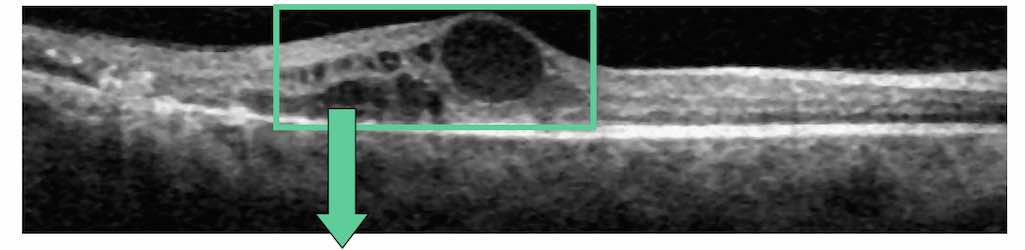
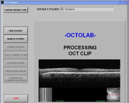
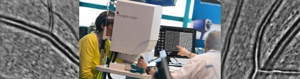
The Laboratory is involved in the assessment of the efficiency and efficacy of a recently developed non invasive diagnostic instrument, able to provide high-quality images of the retina, by means of adaptive optics. In collaboration with the Chair of Internal Medicine of the University (Prof. D. Rizzoni), the instrument and its software are tested for accuracy and repeatability on artificial vessel models, and on a database of subjects.

Based on the data collected so far, the instrument performs in a quite satisfactory way as compared to the previously used techniques.

Related publications

Ciuceis, C.; Caletti, S.; Coschignano, M. A.; Rossini, C.; Duse, S.; Docchio, F.; Pasinetti, S.; Zambonardi, F.; Semeraro, F.; Sansoni, G.; Agabiti Rosei, C.; Pileri, P.; Agabiti Rosei, E.; Rizzoni, D. “Comparison between techniques of evaluation microvascular morphology: The gold-standard locally invasive micromyography vs. three non-invasive techniques. Preliminary data.” Artery Research, Vol 20, pp 95-96. 2017

Rizzoni, D.; Agabiti Rosei, C.; De Ciuceis, C.; Semeraro, F.; Rizzoni, M.; Docchio, F. “New Methods to Study the Microcirculation“, American Journal of Hypertension, Vol 31, Issue 3, pp 265–273. 2018

Nardin, M.; Coschignano, M. A.; Rossini, C.; De Ciuceis, C.; Caletti, S.; Rizzoni, M.; Docchio, F.; Porteri, E.; Rizzoni, D. Methods of evaluation of microvascular structure: state of the art“, European Journal of Translational and Clinical Medicine, Vol 1, pp 7-17. 2018

The OCT-MAT Project: early diagnosis of ocular edemas

The aim of this project was to develop a practical, user friendly software tool in order to have significant and quantitative information on macular edema from OCT images. The activity has been carried out in collaboration with the Cattedra di oculistica dell’ospedale di Varese (Italy), in 2006-2008. We have developed a novel, dedicated software, called OCT-Measurements Analysis Tool (OCT-MAT), that acquires the avi files from an OCT 3 (Carl Zeiss, Meditec, Dublin, CA, USA) and a OCT/SLO (Ophthalmic Technologies Inc., Toronto, Canada) instruments, and carries out five subsequent elaboration steps. These are (i) the creation of patient’s folder, (ii) the selection of frames from the avi files, (iii) the filtering of the frames, (iv) the extraction of quantative information from the hyporeflective areas and their analysis, and (v) the comparison among frames for patient follow-up.

The OCT-MAT software is able to process and output quantitative information in terms of the number of empty spaces and of their extension in micron2. In addition, it allows us to analyze edema reabsorption during time, by suitable clustering of macular empty spaces (i.e., hyporeflective areas). This new OCT-MAT software has been applied on 30 healthy eyes and on three groups of patients with different macular diseases.The use of the OCT-MAT software allows us to measure the edema in terms of both the number of empty spaces and of their extension directly from the inside retina. Suitable tools are available to customize the standard modalities of operation, in order to optimize the measurements. A dedicated staging system gives a quick interpretation of the results.

The possibility of comparing macula edema measurements belonging to different images processed in subsequent times results in the achievement of numerical information, instead of simple qualitative evaluation, on the evolution of macular edema as well as on edema clearance. The OCT-MAT can be installed on standard, non-dedicated hardware, and the learning curve of an experienced retina specialist is very short.

oct_front_panel.jpg

Relevant Publications

Sansoni, G.; Tironi, M.; Trebeschi, M.; Tottoli, F.; Azzolini, C.; Donati, S. “Diagnostica oculare mediante sistema di analisi per la misura di aree iporiflettenti intraretiniche“, Atti del XXIV Congresso Nazionale Associazione “Gruppo di Misure Elettriche ed Elettroniche”, pp. 319-320. 2007

Prati, M.; Donati, S.; Tartaglia, V.; Sansoni, G.; Tironi, M.; Chelazzi, P.; Brancato, R.; Azzolini, C. “Correlation Between Visual Acuity and Retinal Sensitivity Before and After Surgery for Macular Diseases“, Investigative Ophthalmology and Visual Sciences, Vol. 49, no. 13. 2008

Donati, S.; Sansoni, G.; Tironi, M.; Chelazzi, P.; Brancato, R.; Azzolini, C. “Evaluation of results of macular surgery: Role of microperimetry-related OCT imaging study“, Abstract and Presentation, 8th. EURETINA congress, Vienna. 2008

Wavelet image analysis

The main targets of the project are (a) medical images and (b) biological images. For medical images we would develop new image manipulation and analysis tools, specifically designed for noise detection, image segmentation and 3D visualization on a real time, internet shared platform. For biological images, we would develop image downloading, deconvolution and uploading algorithms specifically designed to extract measurement information from compressed stacks of images over the web in real time.

While software currently exists to do these types of manipulations on medical images and (to a lesser extent) on biological images, it is not easy to use, has an enormous learning curve, is very expensive, and is single-user based in its operation. Our success will open up a very exciting opportunity to make a significant change in the field, bringing image analysis capabilities to many more users and at a cost far below the current price.

The work performed up to now has led to the development of wavelet-based algorithms aimed at fast, transparent, automatic denoising of Gaussian and Poisson affected images, taken from both Wide-Field and Confocal microscopes. We chose the wavelet paradigm because of (i) its ability to elaborate sparse images for denoising, and (ii) its ability to perform image compression. Both denoising and compression represent two key-aspects as far as web-based image transmission, elaboration and multi-resolution are concerned. In addition, we thought strategic to implement the DWT algorithm on the FPGA hardware, to improve efficiency and to make it possible to embed the transformation on a hardware independent platform.

The MICE Project

This activity has been accomplished in collaboration with the dept. of Physics of the Politecnico of Milan. The objective was to develop and test specific image processing algorithms based on piece-wise linear histogram transformation to assist tumor detection by means of a time-gated fluorescence imaging technique. The developed procedures have been designed to improve, in real time, the quality of the images taken by means of an intensified video camera. Smart optimization criteria have been followed for the automatic choice of the enhancement parameters. An example applied to the detection of experimental tumors induced in mice is shown in the figures below.

Development of a WEB-based image analysis service for biological and medical images

The aim of this project is to develop and test novel image processing algorithms for image deconvolution and analysis specifically designed for real time, hardware-independent operations. The final goal is to share the image analysis capabilities using cloud computing techniques, by means of a specifically developed web-based system.

This project involves a lasting collaboration between the faculty of Engineering at Università degli Studi di Brescia and the Mechanical Engineering department at MIT. It is supported by the CARIPLO UniBS-MIT-MechE project, a  faculty exchange program agreement designed to promote new long term scientific collaborations and to consolidate existing collaborations between faculty members of UniBS and MIT-MechE.

The faculties involved into this program are Prof. Giovanna Sansoni, for the University of Brescia, and Prof. Forbes Dewey, for the Massachusetts Institute of Technology.

As part of project, Prof. Sansoni of the University of Brescia visited MIT for three months in the early Fall of 2011 to inaugurate the personal exchange program. Prof. Sansoni gave a lecture at MIT, entitled “3D imaging and surroundings“.

Dewey_sansoni
Prof. Giovanna Sansoni's seminar "3D imaging and surroundings"

During her stay at MIT, she had the opportunity to meet a number of Prof. Dewey’s collegues. Among them, Prof. George Barbastiathis gave a number of useful suggestions on non-linear image analysis approaches. The exchange she had at MIT made it possible to establish the basics for an extremely stimulating research field, dealing with image denoising, deconvolution and compression to be performed across the web.

Prof. Dewey travelled to Brescia for two weeks in April of 2012. The aim of his visit was twofold: on one hand, he interacted with Prof. Sansoni Laboratory to optimize the collaboration. He also met people from the Laboratory Start-Ups, and realized how rich and powerful is the high-technology pole generated by the Laboratory.

On the other hand, he gave two seminars open to Ph. D. students and researchers of the University of Brescia. In the first seminar, entitled “Quantitative Models of Cellular Biology: Application to Cellular Dynamics and Drug Development“, he gave two examples of how one can begin to put large-scale quantitative biological models together to predict ab into the results of altering drug dosages and creating drug combinations. In the second seminar, enitled “Super-Resolution Optical Microscopy: Beating Photons at Their Own Game“, he addressed the application of measuring the mechanical properties of living cells using 3D stacks from optical microscope images, far beyond the Rayleigh criteria.

Prof. Dewey's seminar "Application to Cellular Dynamics and Drug Development"
Prof. Dewey’s Seminar “Super-Resolution Optical Microscopy: Beating Photons at Their Own Game”
Prof. Dewey and Prof. Sansoni with Prof. Beretta, the director of the CARIPLO-MIT program