Wavelet image analysis

The main targets of the project are (a) medical images and (b) biological images. For medical images we would develop new image manipulation and analysis tools, specifically designed for noise detection, image segmentation and 3D visualization on a real time, internet shared platform. For biological images, we would develop image downloading, deconvolution and uploading algorithms specifically designed to extract measurement information from compressed stacks of images over the web in real time.

While software currently exists to do these types of manipulations on medical images and (to a lesser extent) on biological images, it is not easy to use, has an enormous learning curve, is very expensive, and is single-user based in its operation. Our success will open up a very exciting opportunity to make a significant change in the field, bringing image analysis capabilities to many more users and at a cost far below the current price.

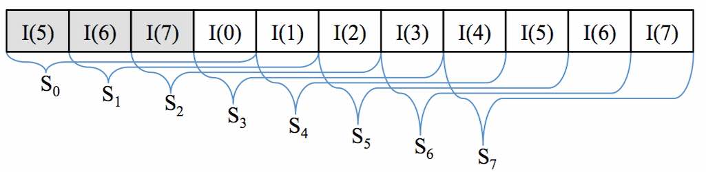
The work performed up to now has led to the development of wavelet-based algorithms aimed at fast, transparent, automatic denoising of Gaussian and Poisson affected images, taken from both Wide-Field and Confocal microscopes. We chose the wavelet paradigm because of (i) its ability to elaborate sparse images for denoising, and (ii) its ability to perform image compression. Both denoising and compression represent two key-aspects as far as web-based image transmission, elaboration and multi-resolution are concerned. In addition, we thought strategic to implement the DWT algorithm on the FPGA hardware, to improve efficiency and to make it possible to embed the transformation on a hardware independent platform.

The MICE Project

This activity has been accomplished in collaboration with the dept. of Physics of the Politecnico of Milan. The objective was to develop and test specific image processing algorithms based on piece-wise linear histogram transformation to assist tumor detection by means of a time-gated fluorescence imaging technique. The developed procedures have been designed to improve, in real time, the quality of the images taken by means of an intensified video camera. Smart optimization criteria have been followed for the automatic choice of the enhancement parameters. An example applied to the detection of experimental tumors induced in mice is shown in the figures below.

Development of a WEB-based image analysis service for biological and medical images

The aim of this project is to develop and test novel image processing algorithms for image deconvolution and analysis specifically designed for real time, hardware-independent operations. The final goal is to share the image analysis capabilities using cloud computing techniques, by means of a specifically developed web-based system.

This project involves a lasting collaboration between the faculty of Engineering at Università degli Studi di Brescia and the Mechanical Engineering department at MIT. It is supported by the CARIPLO UniBS-MIT-MechE project, a  faculty exchange program agreement designed to promote new long term scientific collaborations and to consolidate existing collaborations between faculty members of UniBS and MIT-MechE.

The faculties involved into this program are Prof. Giovanna Sansoni, for the University of Brescia, and Prof. Forbes Dewey, for the Massachusetts Institute of Technology.

As part of project, Prof. Sansoni of the University of Brescia visited MIT for three months in the early Fall of 2011 to inaugurate the personal exchange program. Prof. Sansoni gave a lecture at MIT, entitled “3D imaging and surroundings“.

Dewey_sansoni
Prof. Giovanna Sansoni's seminar "3D imaging and surroundings"

During her stay at MIT, she had the opportunity to meet a number of Prof. Dewey’s collegues. Among them, Prof. George Barbastiathis gave a number of useful suggestions on non-linear image analysis approaches. The exchange she had at MIT made it possible to establish the basics for an extremely stimulating research field, dealing with image denoising, deconvolution and compression to be performed across the web.

Prof. Dewey travelled to Brescia for two weeks in April of 2012. The aim of his visit was twofold: on one hand, he interacted with Prof. Sansoni Laboratory to optimize the collaboration. He also met people from the Laboratory Start-Ups, and realized how rich and powerful is the high-technology pole generated by the Laboratory.

On the other hand, he gave two seminars open to Ph. D. students and researchers of the University of Brescia. In the first seminar, entitled “Quantitative Models of Cellular Biology: Application to Cellular Dynamics and Drug Development“, he gave two examples of how one can begin to put large-scale quantitative biological models together to predict ab into the results of altering drug dosages and creating drug combinations. In the second seminar, enitled “Super-Resolution Optical Microscopy: Beating Photons at Their Own Game“, he addressed the application of measuring the mechanical properties of living cells using 3D stacks from optical microscope images, far beyond the Rayleigh criteria.

Prof. Dewey's seminar "Application to Cellular Dynamics and Drug Development"
Prof. Dewey’s Seminar “Super-Resolution Optical Microscopy: Beating Photons at Their Own Game”
Prof. Dewey and Prof. Sansoni with Prof. Beretta, the director of the CARIPLO-MIT program